


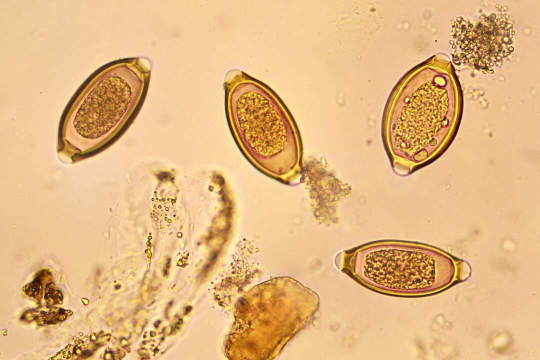
In this article we will try to understand what the most common symptoms of helminthiasis in adults are, whether they can be asymptomatic and also which types of helminth infestations are most widespread in our country.
Introduction to terminology
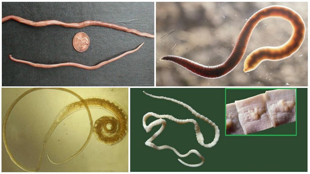
Human helminth infections are usually caused by parasitic worms of three classes: Nematoda (roundworms) - roundworms, Cestoda (tapeworms) - tapeworms, Trematoda (trematodes) - trematodes.According to the class of the pathogen, all human helminthiases are conventionally divided into:
- Roundworms are helminth infections associated with infestation with roundworms or their larvae.The best known nematodes are enterobiasis, ascariasis, hookworm, toxocariasis and strongyloidosis.
- Cestodoses are caused by the parasitism of tapeworms or their larval forms.The most common cestodiasis are diphyllobothriasis, taeniasis and teniarrhiasis, hymenolepiasis and echinococcosis.
- Flukes are helminth infections caused by different types of flukes.The most widespread diseases in our country are opisthorchiasis, clonorchiasis, less commonly fascioliasis and very rarely paragonimiasis.
In relation to the territory of our country, all helminthiases can be divided into:
- Typical, that is, often found among the population of our country;
- Exotic, caused by population migration and tourism to other countries.
Detection and treatment of exotic helminth infections is a more complex and time-consuming process, as laboratories may not have the necessary reagents to perform laboratory tests.The following types of helminth infestations are typical of the territory of our country.
Which population groups are most susceptible to infections?
Helminth infestations occur most often in the following groups:
- Young children due to active knowledge of the world and lack of personal hygiene skills.
- Children organized into groups (nurseries, schools, camps, sanatoriums, etc.).Close interaction, shared habitat and toy exchange contribute to the spread of geohelminthiasis with a simple life cycle.
- Adults who have close contact and work with children, including teachers and parents.
- People with certain eating habits: consumption of raw or minimally processed meat, fish, seafood, aromatic herbs, etc.Great importance is attached to national cuisine (sushi, stroganina, yukola, porsa and other dishes), love of drying, salting and smoking.
- People living in endemic, tropical and subtropical areas.
- People living in poor sanitation conditions, with lack of access to quality water resources and sanitation services.
- People closely linked to agriculture and livestock raising, who live close to animals.
When and who should get tested?
Based on belonging to risk groups and the characteristics of the course of helminth infestations, the examination for helminthiasis should be performed in the following cases:
- Children, organized and unorganized, at least once a year in the absence of symptoms.
- Adults working with children and in catering establishments, doctors, sellers of children's items and food products.
- Children and adults entering educational institutions, sanatoriums, hospitals, boarding houses, hospices and other institutions that involve the presence of a large number of people in one area.
- Children and adults with allergic skin and bronchopulmonary diseases resistant to traditional therapeutic methods and elimination diets.
- Children and adults with symptoms of chronic damage to the gastrointestinal tract: nausea, vomiting, widespread abdominal pain, diarrhea, pain in the epigastric and periumbilical area, malabsorption syndrome.
- Children and adults with symptoms of damage to the hepatobiliary system - hepatosplenomegaly, jaundice, pain in the right hypochondrium, increased AST, ALT, GGTP, alkaline phosphatase, portal hypertension.
- Children and adults affected by mild fever of unknown etiology for a long time.
- Children and adults with severe asthenic syndrome: frequent headaches, weight loss, bulimia or lack of appetite, unexplained weakness, fatigue, sleep disturbances.
- Children and adults with microcytic and normocytic anemia resistant to traditional treatment.Vitamin B12 deficiency anemia accompanies 2-4% of cases of diphyllobothriasis, less often teniarrhiasis, ascariasis and other helminthiasis.
Characteristics of the course of helminth infestations
The course of any helminth infestation can be divided into several periods:
- Infection and its clinical manifestations.Most helminth infections have no symptoms of infection, but with strongyloidiasis, hookworm and some others, so-called cercariasis occur - diseases caused by the penetration of the larvae into the skin.
- The incubation period is the time from the moment of infection to the first clinical manifestations.
- The acute phase, most often caused by the migration of larvae into the human body, their moulting, maturation into sexually mature individuals, first egg-laying and the first encounter of the human body with unknown antigens.The duration of the acute phase usually varies from several days to several weeks.Sometimes the acute phase is absent or weakly expressed, which is more often observed with a low degree of invasion or in indigenous populations who have “immune memory”.
- The chronic phase occurs in the absence of treatment or ineffectiveness and is associated with the direct influence of sexually mature individuals.
The duration of the infestation depends on the lifespan of the helminths and the likelihood of self-infection.
From this point of view, the course of all helminthiases can be represented in the form of several basic schemes.
- Scheme 1: The course of the disease has the shape of a sinusoid.Onset (time of infection) – increase in symptoms up to a certain level (degree of severity) – transition to the chronic phase with periodic exacerbations.This type of course is characteristic of opisthorchiasis, clonorchiasis, fascioliasis and strongyloidosis.
- Scheme 2: The course of helminthiasis has the form of a plateau.Infection with subsequent increase in symptoms up to a certain point, after which the patient notices that the symptoms remain constant or decrease and disappear.A similar course is typical of many invasions: trichinosis, taeniasis, diphyllobothriasis, ascariasis.
- Scheme 3: the course of helminthiasis is characterized by a continuous increase in symptoms, which can ultimately lead to the death of the patient.These are, as a rule, massive invasions or helminthiasis, occurring with diffusion against the background of immunodeficiency (disseminated strongyloidosis).
Next, let's look at where worms live in the human body.Helminth infections can affect any human organ, however, by systematization, several main "preferred" localizations in the host's body can be identified:
- The intestine is the most logical habitat for many human worms, for example, roundworms, pinworms, whipworms, hookworms, bovine and porcine tapeworms, etc.Here the parasite attaches itself in one of the ways to the intestinal wall and carries out its "subversive" activity, most often stealing nutrients from the host's food.
- The liver and bile ducts are the preferred habitat of opisthorchids, clonorchids and flukes.In addition, the liver is often affected during the migration of helminth larvae, for example, lung fluke, echinococcus, etc.
- Lungs and bronchi.The life cycle of many nematodes (roundworms, hookworms, intestinal eels) cannot proceed without the migration of the larvae through the bronchopulmonary system.For other helminths, the lungs are the final target and habitat of adult individuals (paragonimiasis).
- Central nervous system.It is most often affected by cysticercosis, echinococcosis, toxocariasis, paragonimiasis and other invasions.This is usually due to the migration of the larval forms and their spread through the bloodstream.
- The skin, as a rule, becomes the habitat of larval forms and various filariae.
- Eyes: filariasis, toxocariasis.
- Venous plexuses of the bladder, uterus and intestine in schistosomiasis.
Symptoms of helminthiasis
In general, the clinical picture of helminth infestations consists of the following syndromes:
- Toxic-allergic.The main reasons for the development of allergic and toxic reactions are the metabolic products of adult individuals and larval forms and the release of toxins by them.Worms in humans can cause skin rashes such as hives, allergic and atopic dermatitis, and sometimes eczema.Skin rashes have a recurrent course and rarely respond to traditional treatment.
- Dyspeptic.Dyspeptic symptoms include nausea, vomiting, heartburn, belching, bitterness in the mouth, and flatulence.Diarrhea or loose stools without increased frequency of bowel movements are common.Dyspepsia is more typical for helminths living in the intestine (ascariasis, trichuriasis, etc.) and rarely occurs with tissue helminthiasis (echinococcosis, cysticercosis, toxocariasis, etc.).
- Indigestion and malabsorption syndrome.It is also more typical of intestinal invasions, especially those in which sexually mature parasites live in the small intestine and duodenum (taeniasis, diphyllobothriasis, etc.)
- Abdominal pain: in children - more often widespread, in the umbilical region, in the right iliac region, simulating appendicitis.In adults, the localization of pain is more specific.Sometimes the pain syndrome due to helminth infestations can simulate an “acute abdomen”.
- Bronchopulmonary.In most cases it is caused by the migration of the parasite larvae into the alveoli, followed by movement into the bronchial tree, trachea and oropharynx.The main symptoms at this stage are cough, usually productive, with mucous or mucopurulent sputum, the appearance of wet and dry wheezing, a change in the nature of breathing to harsh or harsh with prolongation of exhalation.With such invasions (ascariasis, strongyloidosis, hookworm), Loeffler syndrome can be observed.In some invasions (for example in paragonimiasis), the lungs are the usual habitat of adult individuals.The clinical manifestations in this case are different.Chronic cough with sputum and blood, hemoptysis, radiological changes such as pneumofibrosis, cystic lesions of the lung tissue often imitate the picture of pulmonary tuberculosis.
- Worms in humans can cause anemic syndrome.The development of anemia can be caused by direct consumption of the host's blood (hookworm), host robbing (ascariasis, taeniasis, diphyllobothriasis, etc.), dysfunction of the stomach and intestines (taeniarinosis, taeniasis, etc.), and exposure to toxins.The anemia is most often characterized by microcytic iron deficiency, and in some helminthiases it is macrocytic and B-12 deficient.
- Asthenia, as a sign of the presence of worms in the human body, is characterized by sleep disorders, dyssomnia, increased irritability, aggression, increased fatigue, hyperactivity and attention deficit in children.It is difficult for the patient to concentrate on work and concentrate.An infected person may experience unexplained weight loss, loss of appetite (or, conversely, increased appetite).
- Intoxication-inflammatory syndrome manifests itself in the form of an increase in temperature up to 38 degrees and above, myalgia and arthralgia, leukocytosis and increased ESR in the CBC.The degree of its severity depends on the patient's immune status and the stage of invasion.The acute phase occurs more often than the chronic one with fever and flu-like condition.The chronic phase is more characterized by low-grade fever or normal body temperature.
- Worms in humans are often the cause of unexplained asymptomatic eosinophilia.Eosinophilia is more characteristic of roundworms and trematodes and may be accompanied by general leukocytosis.
In the following table we have tried to summarize the information available on the symptoms of worm infection that accompany the most common helminth infestations in humans.
| Helminthiasis | Symptoms of infection | Incubation period | Acute phase | Chronic phase |
|---|---|---|---|---|
| Enterobiasis | No | 10-15 days | Perianal itching, diffuse abdominal pain, rarely diarrhea, nausea, vomiting.Dyssomnia, nighttime screaming and crying in children.Persistent vulvovaginitis, lip synechiae. The total duration of the invasion is about a month or two (if there is no reinfection) |
|
| Ascariasis | No | About a week | Up to 2 weeks, the acute phase is caused by the migration of the larvae.Symptoms during this period are fever, muscle and joint pain, skin rashes and itching, signs of bronchitis or pneumonia (eosinophilic infiltrative changes in the lung tissue).Bronchoobstruction may occur. | Decreased appetite, nausea, vomiting, abdominal pain (near the navel, right iliac region), rumbling in the intestines and bloating.Symptoms of asthenia are persistent headaches, weakness, fatigue.Symptoms of anemia and hypovitaminosis.Total duration of invasion (in the absence of reinfection) - 1 year |
| Trichocephalosis | No | 1-1.5 months | Pain along the large intestine, right iliac region, nausea, vomiting, salivation, flatulence, stool instability, presence of blood and mucus in the stool (total duration of invasion is up to 5-7 years). | |
| Diphyllobothriasis | No | Canceled, as the clinical picture of the invasion develops gradually | There is no acute phase as such.Symptoms appear and progress slowly.General asthenia, which increases over time, skin rashes, diffuse abdominal pain, sometimes in the right iliac region, nausea, vomiting, loss of appetite and progressive weight loss, stool instability, signs of anemia, glossitis, hepatosplenomegaly, neurological symptoms (paraesthesia, impaired sensitivity and motor function, etc.). The duration of the invasion reaches up to 10 years or more. |
|
| Taeniasis and teniarinhoz | No | Canceled, as the clinical picture of the invasion develops gradually | There is no acute phase as such.The following symptoms appear and increase: weakness, fatigue, headache and other symptoms of asthenia, mild or moderate anemia, widespread abdominal pain and discomfort, bulimia or loss of appetite, progressive weight loss, drooling, nausea, vomiting, skin rashes such as urticaria, rarely damage to the nervous system such as Meniere's syndrome, epileptiform seizures. | |
| Opisthorchiasis | No | 2-4 weeks | Increased temperature, weakness, fatigue, eosinophilia and leukocytosis, pain in the abdomen and liver, loose stools, allergic rash, hepatosplenomegaly, jaundice. | Symptoms of intestinal and hepatobiliary dyspepsia, hepatomegaly, jaundice, pain in the liver, there may be biliary colic, pain in the girdle (pancreatitis), a gradual increase in liver failure. |
| Clonorchiasis | No | 2-3 weeks | The same as opisthorchiasis, but more pronounced | The same as for opisthorchiasis.High probability of developing cirrhosis and cholangiocarcinoma |
| Hookworms and necatoriasis | 2-3 days after infection, an itchy papular or urticarial rash appears at the site of penetration of the larvae, often resembling tortuous passages.The duration of this period is up to 1.5 - 2 weeks. | Virtually absent | Due to the migration of the larvae, approximately 2-4 weeks.Fever, skin rashes, symptoms of bronchitis, bronchopneumonia - productive cough, wet and dry wheezing, sometimes bronchial obstruction.Characteristic is the formation of volatile eosinophilic infiltrates in the lung tissue (Leffler syndrome). | The most important clinical sign is moderate to severe iron deficiency anemia.The second significant criterion is hypoalbuminemia, edema syndrome.Nausea, vomiting, pain in the epigastric region, flatulence, fatigue, headache, general asthenia.The total duration of the invasion is up to 20 years. |
| Hymenolepiasis | No | It is expressed implicitly, for the maturation of an adult from a larva 2-3 weeks are enough | Therefore, the acute phase is not pronounced;the symptoms increase and are characterized by nausea, vomiting, salivation, widespread abdominal pain and loose stools.Damage to the nervous system in the form of headaches, dizziness, fainting.Allergic manifestations: exanthema-like skin rash, urticaria, Quincke's edema, allergic rhinitis.Pale skin with a jaundiced tinge.Body temperature is often normal. | |
| Fascioliasis | No | 1-8 weeks | Fever, liver and epigastrium pain, jaundice, hepatomegaly, loose stools, allergic rash, eosinophilia, and leukocytosis | Hepatomegaly, liver pain, hepatobiliary dyspepsia, increased hepatic failure, secondary cholecystitis and pancreatitis |
| Strongyloidosis | On days 1-2, a slight itching and papular rash appear at the site of penetration of the larvae, which quickly disappear. | Virtually absent | On the 3-4th day from the moment of infection, fever, myalgia, joint pain, cough with sputum, wet and dry wheezing occur, and bronchial obstruction is possible.X-ray reveals volatile eosinophilic infiltrates (Leffler syndrome).Often an allergic rash appears on the skin. | Pain in the epigastric, periumbilical areas, less often in the liver, belching, heartburn, nausea, vomiting, loss of appetite and weight, biliary dyspepsia, diarrhea (usually enteritis type, impurities in the stool are rare, usually with massive invasion), hepatomegaly, jaundice.The infestation lasts for years due to regular self-infection. |
| Trichinosis | Abdominal pain, nausea, vomiting, diarrhea may occur | From 1-2 days to 4-5 weeks, average 10-25 days | After infection, the intestinal phase begins first - abdominal pain, nausea, diarrhea, then generalized - severe muscle pain, swelling of the eyelids, face, less often of the torso, rash depending on the type rash, in more severe cases hemorrhagic, high fever (sometimes mild fever) for several weeks. |
Encapsulation of larvae and their retention in skeletal muscles for 10 years or more. |
| Paragonimiasis | No | 2-3 weeks, can be shortened to several days | Abdominal pain, acute abdominal pain, diarrhoea.Gastroenterological symptoms are replaced by symptoms of damage to the bronchi and lung tissue - cough with sputum, chest pain, pneumonia, exudative pleurisy | Prolonged fever or low-grade fever, prolonged cough with sputum and blood, hemoptysis, chest pain, shortness of breath, weight loss, x-ray shows pulmonary fibrosis, pleural adhesions, thin-walled cysts in the lower parts of the lungs |
How to confirm the presence of worms in the body?
We have arrived at the most frequently asked questions when visiting a doctor.How can you tell if there are worms in the human body?Is it possible to do it yourself without leaving home?
So, it is possible to determine the presence of a helminth infestation in the house only in the following cases:
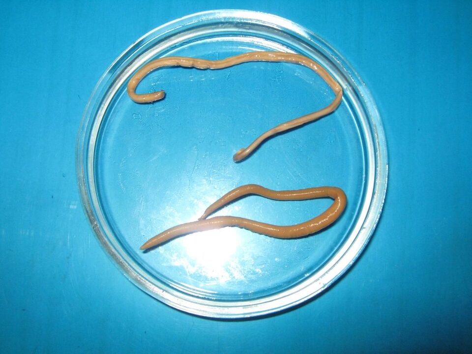
- Visualization of worms in feces (live or dead adult helminths, their fragments).Of course, only helminths that live in the human intestine can be found in feces.As a rule, helminths are detected during a massive infestation.
- Visualization of tapeworm segments in stool.
In all other cases, the diagnosis can be confirmed using 2 main laboratory diagnostic methods:
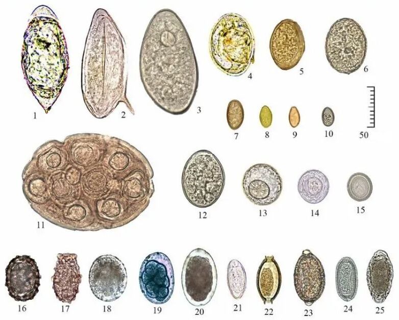
- Various modifications of ovoscopy.Worm eggs have morphological differences and microscopic dimensions;trying to examine them in the feces is useless.
- Serological reactions, including PCR.

For different invasions, the range of laboratory tests is different.It is important to understand that standard analysis of feces for helminth eggs and scraping for enterobiasis are screening methods aimed at identifying the most common intestinal helminth infections in risk groups.At the same time, single microscopy provides only about 50% of information.You can see what worm eggs look like under a microscope in the following figure.The vertical scale represents the size in µm.
Drug therapy and chemoprophylaxis
Treatment of human helminthiasis involves the prescription of appropriate, as well as symptomatic and pathogenetic, anthelmintic agents.In this article we will not consider in detail the doses and treatment regimens for individual helminth infestations;we will only note that the prescription of drugs should be carried out by a doctor (pediatrician, therapist, infectious disease specialist, parasitologist) in accordance with the clinical picture, laboratory test data and the patient's condition.

Helminthiasis chemoprophylaxis is the prophylactic administration of anthelmintic drugs active against geohelminthic infections in risk groups and endemic areas.Since the overall incidence of geohelminthiasis in the entire population is low, chemoprophylaxis can be performed once a year, preferably in autumn or spring.
Preventive measures
Measures to prevent infection with worms are conventionally divided into individual and public.Individual prevention includes:
- Respect for personal hygiene rules among family members and in groups, teaching children the rules and monitoring their implementation.
- Careful processing of vegetables, greens and fruits before direct consumption.
- I refuse to consume raw and undercooked meat of pigs, cattle and other animals, salted, dried and smoked meat that has not passed sanitary and epidemiological checks.
- I refuse to consume fish that has not passed sanitary and epidemiological control, including salted, smoked, dried, caviar and other fish products.
- Drink only high-quality water for drinking and cooking, even when you travel.
- Before leaving for tropical and subtropical countries it is necessary to obtain advice from an infectious disease specialist (parasitologist) and recommendations for laboratory diagnostics after return.The infectious disease specialist also decides on the need for chemoprophylaxis.
- Refusal to swim in fresh water, walk barefoot, or lie on grass in endemic tropical countries.
- I refuse to eat food on the streets, unverified bars and other public catering establishments.
- Annual clinical examination with TAO, OAM, standard coproovoscopy and curettage for enterobiasis.These events are especially relevant in families with children, adults working in organized children's groups, in hospitals, sanatoriums, etc.
Public prevention consists of organizing control of food products on sale in shops and markets, timely identification of sick domestic animals and infected people and their treatment.Of great importance is the monitoring of water resources, the improvement of populated areas, the assessment of the degree of infection of wild mammals, the control of the borders of natural focal helminthiases and the number of stray animals.